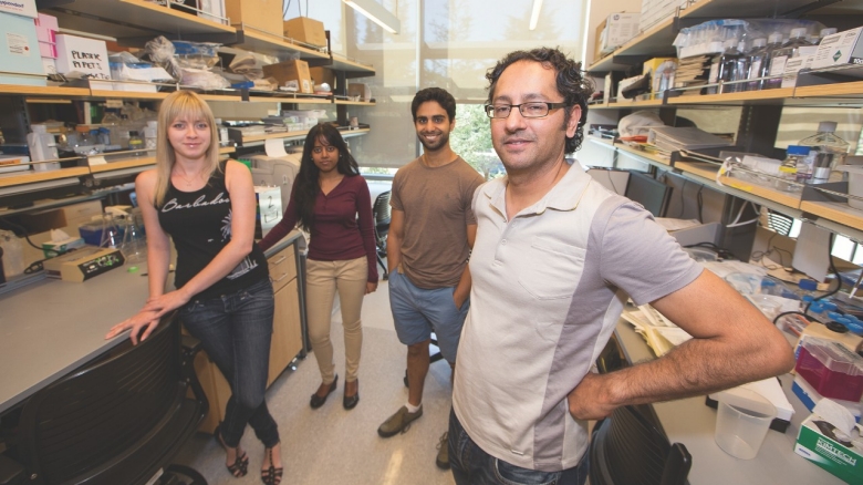
From left: Monica Nagendran, Yana Kazadaeva, and Ahmad Nabhan, with Tushar Desai, MD, MPH
Painting a New Picture of Lung Development

From left: Monica Nagendran, Yana Kazadaeva, and Ahmad Nabhan, with Tushar Desai, MD, MPH
Painting a New Picture of Lung Development
From the outside, the lungs develop like the roots of a plant; branching airways expand and grow increasingly more intricate, until they’ve filled every space they can with ever smaller passageways to capture oxygen from a breath of air. But inside the cells that make up these airways, an even more amazing molecular dance is taking place, one that creates new lung cells—as an embryo develops and, later, in adult lungs. It’s only in the past few years that researchers have begun to understand the details of this story, thanks to an interdisciplinary team at Stanford. And what they’re finding may allow clinicians to learn how to repair lungs in patients with conditions like emphysema and pulmonary fibrosis, or even treat lung cancer.
“You can take intermittent snapshots of what a tissue looks like as it develops, but to really understand it, you want to know what’s happening at a molecular scale between those snapshots,” says Stanford pulmonologist Tushar Desai, MD, MPH (assistant professor, Pulmonary and Critical Care). “To reveal that molecular level of development, though, is very painstaking, time consuming, and hard to get people excited about.”
Most researchers, he said, have skipped from the visual snapshots of lung development to genetic experiments. By engineering mice to lack certain genes, and then studying the effect on the lungs, they can elucidate what genes and molecules are key to the process.
But that’s not the same thing, Desai argues, as understanding each sequential event in lung formation. So, after a medical fellowship in pulmonology and then a post-doctoral research fellowship in the lab of Stanford biochemist Mark Krasnow, MD, PhD (professor, Biochemistry), Desai made it his goal to paint a new, more detailed, picture of how lungs develop.
Inside each tiny airsac of the lung, two types of cells help the body breathe air. Alveolar type 1 (AT1) cells lie flat on the surface of each airsac, enabling the exchange of carbon dioxide and oxygen. Chunkier alveolar type 2 (AT2) cells studding the walls and ceilings produce surfactant, a fluid that coats the airsacs and keeps them from collapsing.
From the outside, the lungs develop like the roots of a plant; branching airways expand and grow increasingly more intricate, until they’ve filled every space they can with ever smaller passageways to capture oxygen from a breath of air. But inside the cells that make up these airways, an even more amazing molecular dance is taking place, one that creates new lung cells—as an embryo develops and, later, in adult lungs. It’s only in the past few years that researchers have begun to understand the details of this story, thanks to an interdisciplinary team at Stanford. And what they’re finding may allow clinicians to learn how to repair lungs in patients with conditions like emphysema and pulmonary fibrosis, or even treat lung cancer.
“You can take intermittent snapshots of what a tissue looks like as it develops, but to really understand it, you want to know what’s happening at a molecular scale between those snapshots,” says Stanford pulmonologist Tushar Desai, MD, MPH (assistant professor, Pulmonary and Critical Care). “To reveal that molecular level of development, though, is very painstaking, time consuming, and hard to get people excited about.”
Most researchers, he said, have skipped from the visual snapshots of lung development to genetic experiments. By engineering mice to lack certain genes, and then studying the effect on the lungs, they can elucidate what genes and molecules are key to the process. But that’s not the same thing, Desai argues, as understanding each sequential event in lung formation. So, after a medical fellowship in pulmonology and then a post-doctoral research fellowship in the lab of Stanford biochemist Mark Krasnow, MD, PhD (professor, Biochemistry), Desai made it his goal to paint a new, more detailed, picture of how lungs develop.
Inside each tiny airsac of the lung, two types of cells help the body breathe air. Alveolar type 1 (AT1) cells lie flat on the surface of each airsac, enabling the exchange of carbon dioxide and oxygen. Chunkier alveolar type 2 (AT2) cells studding the walls and ceilings produce surfactant, a fluid that coats the airsacs and keeps them from collapsing.
Scientists had previously hypothesized that progenitor cells in the developing lungs acted as the precursors for AT2 cells, and that some AT2 cells could then form from AT1 cells. But when Desai, in collaboration with Krasnow, traced the origin of lung cells, he discovered that a single alveolar progenitor cell directly formed both cell types. In the lungs of adults, however, these precursor cells were nowhere to be found. Instead, some AT2 cells acted as stem cells—able to form both new AT2 and AT1 cells. The results were published last year in the journal Nature.
“One of the most surprising things was that rare AT2 cells seem to be bifunctional,” says Desai. “Not only are they acting as stem cells, but they’re also apparently still secreting surfactant and keeping the lung functional that way.”
Desai went on to capture the transcriptome—levels of genes being used by a cell—in precursor and newly forming AT1 and AT2 cells. By determining what genes are turned on and off during this dynamic process, he thinks he may be able to find the molecular switch that’s flipped to generate new AT1 and AT2 cells.
Answering that question, Desai says, will not only satisfy his quest for understanding lung development, but could lead to new therapeutics for lung diseases.
